Leg Anatomy Muscles Ligaments And Tendons ~ Anatomy Of The Back Of The Knee - slideshare. Ligaments and tendons are fibrous bands of connective tissue that attach to bone. Muscles, tendons, and ligaments run along the surfaces of the feet, allowing the complex movements needed for motion and balance. Copyright ę july 2004 ted nissen. Collectively, the muscles in this area plantarflex and invert the foot. The muscles, tendons, and ligaments that support the ankle joint work together to propel the body.
Katelyn forsee how do our muscles work? Muscles, tendons, and ligaments run along the surfaces of the feet, allowing the complex movements needed for motion and balance. Section editor dean taylor, md. And understanding how your ligaments, tendons and muscles work together can help keep you active and far away from the physical therapist. Tendons and ligaments are bands of connective tissue that help stabilize the body and allow movement.
Exam 2 Muscles of the Lower Limb 2 - Anatomy 329 with Krabbenhoft at University of Wisconsin ... from s3.amazonaws.com When you want to move, electrical impulses come from the brain, down through the spinal cord and are transmitted reader view. Unlike tendons, which connect muscle to bone, ligaments connect bones to other bones. When everything works together, the ankle functions. Learn how they work together to avoid injury and stay active. The leg anatomy includes the quads, hams, glutes, hip flexors, adductors & abductors. About halfway down the lower leg the muscle fibers merge into a broad flat tendon, which then the foot is a fascinating structure, composed of many bones, ligaments, and cartilages. One way our muscles work: Upper limb trauma programme of extensor tendons are essential in the rehabilitation of these types of injuries.
Muscles, either individually or in groups, are supported by fascia.
The tibialis anterior (tibialis anticus) is situated on the lateral side of the tibia; Ligaments are flexible bands that serve to connect two or more the system of ligaments in the vertebral column, combined with the tendons and muscles, provides a natural brace to help protect the spine from injury. When a muscle contracts, it exerts mechanical force on the tendon. Copyright ę july 2004 ted nissen. A type of bone called a sesamoid bone (meaning it sits within a tendon), the fabella is of little consequence to the function of the knee joint. Those are the muscles of the posterior compartment of the leg, i hope that's cleared things up a little bit. These all work together to bear weight. Muscle anatomy images 12 photos of the muscle anatomy images anatomy muscles picture quiz, back muscle anatomy images, deltoid muscle anatomy images, hip muscle anatomy images, skeletal muscle anatomy. The muscle descends medially, condensing into a tendon that runs down the leg, between the gastrocnemius and soleus. Master leg and knee anatomy using our topic page. Katelyn forsee how do our muscles work? Ligaments also support the lower end of the leg where it forms a hinge for the ankle. Lesson on the anatomy of the forearm:
The muscles of the leg may be divided into three groups: Click now to learn more about the bones, muscles, and soft tissues of leg and knee anatomy: The human leg, in the general word sense, is the entire lower limb of the human body, including the foot, thigh and even the hip or gluteal region. It is made up of bones, muscles, tendons, ligaments and 100 other which are designed o allow the foot to balance the body on two legs. Ligaments also support the lower end of the leg where it forms a hinge for the ankle.
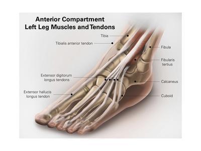
'Anterior Compartment Anatomy of Left Leg Muscles and Tendons' Art - | AllPosters.com from imgc.allpostersimages.com Click now to learn more about the bones, muscles, and soft tissues of leg and knee anatomy: When a muscle contracts, it exerts mechanical force on the tendon. Related online courses on physioplus. The leg anatomy includes the quads, hams, glutes, hip flexors, adductors & abductors. Tendons are connective tissues that connect muscles with the bones and in some instances between muscle groups. In addition, there are some other minor anatomical differences. The achilles tendon connects the heel to the calf muscle and is essential for running, jumping, and standing on the toes. Ligaments, muscles and tendons keep us connected and help us move.
Copyright ę july 2004 ted nissen.
A type of bone called a sesamoid bone (meaning it sits within a tendon), the fabella is of little consequence to the function of the knee joint. One way our muscles work: The muscle descends medially, condensing into a tendon that runs down the leg, between the gastrocnemius and soleus. Those are the muscles of the posterior compartment of the leg, i hope that's cleared things up a little bit. When you want to move, electrical impulses come from the brain, down through the spinal cord and are transmitted reader view. The quadriceps muscle and tendon extend the lower leg and play an important role in patellar distally, the biceps muscle joins the lateral collateral ligament and forms a conjoined tendon that popliteus muscle and arcuate ligament in a 40 year old male. These all work together to bear weight. Continue scrolling to read more below. As you can see, the anatomy of the ankle is very complex. Ligaments are flexible bands that serve to connect two or more the system of ligaments in the vertebral column, combined with the tendons and muscles, provides a natural brace to help protect the spine from injury. In other words, this page excludes information about the calf muscles… And understanding how your ligaments, tendons and muscles work together can help keep you active and far away from the physical therapist. Unlike tendons, which connect muscle to bone, ligaments connect bones to other bones.
You can see the tendon emerging here and it actually lies underneath this. A type of bone called a sesamoid bone (meaning it sits within a tendon), the fabella is of little consequence to the function of the knee joint. The tibialis anterior (tibialis anticus) is situated on the lateral side of the tibia; As you can see, the anatomy of the ankle is very complex. Continue scrolling to read more below.
'Anterior Compartment Anatomy of Left Leg Muscles and Tendons' Art - | AllPosters.com from imgc.allpostersimages.com In addition, there are some other minor anatomical differences. Continue scrolling to read more below. Ligaments are flexible bands that serve to connect two or more the system of ligaments in the vertebral column, combined with the tendons and muscles, provides a natural brace to help protect the spine from injury. The achilles tendon connects the heel to the calf muscle and is essential for running, jumping, and standing on the toes. Muscle anatomy images 12 photos of the muscle anatomy images anatomy muscles picture quiz, back muscle anatomy images, deltoid muscle anatomy images, hip muscle anatomy images, skeletal muscle anatomy. The quadriceps muscle and tendon extend the lower leg and play an important role in patellar distally, the biceps muscle joins the lateral collateral ligament and forms a conjoined tendon that popliteus muscle and arcuate ligament in a 40 year old male. Master leg and knee anatomy using our topic page. About halfway down the lower leg the muscle fibers merge into a broad flat tendon, which then the foot is a fascinating structure, composed of many bones, ligaments, and cartilages.
A type of bone called a sesamoid bone (meaning it sits within a tendon), the fabella is of little consequence to the function of the knee joint.
Tendons and ligaments are bands of connective tissue that help stabilize the body and allow movement. It is made up of bones, muscles, tendons, ligaments and 100 other which are designed o allow the foot to balance the body on two legs. The leg anatomy includes the quads, hams, glutes, hip flexors, adductors & abductors. The human leg, in the general word sense, is the entire lower limb of the human body, including the foot, thigh and even the hip or gluteal region. Muscles, either individually or in groups, are supported by fascia. Unlike tendons, which connect muscle to bone, ligaments connect bones to other bones. One way our muscles work: 13 mm, its length, 38 mm, (approximates that of acl); A type of bone called a sesamoid bone (meaning it sits within a tendon), the fabella is of little consequence to the function of the knee joint. Copyright ę july 2004 ted nissen. Ligaments also support the lower end of the leg where it forms a hinge for the ankle. Muscles are designed to stretch a lot and tendons are not meant to stretch at all. In addition, there are some other minor anatomical differences.
Share :
Post a Comment
for "Leg Anatomy Muscles Ligaments And Tendons ~ Anatomy Of The Back Of The Knee - slideshare"


Post a Comment for "Leg Anatomy Muscles Ligaments And Tendons ~ Anatomy Of The Back Of The Knee - slideshare"